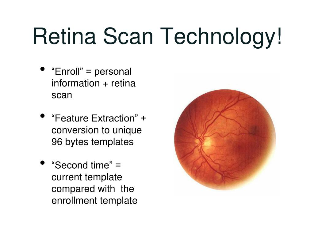

You can't always prevent diabetic retinopathy. Diabetic retinopathy, macular edema, glaucoma or a combination of these conditions can lead to complete vision loss, especially if the conditions are poorly managed. This pressure can damage the nerve that carries images from your eye to your brain (optic nerve). New blood vessels can grow in the front part of your eye (iris) and interfere with the normal flow of fluid out of the eye, causing pressure in the eye to build. This can cause spots floating in your vision, flashes of light or severe vision loss. The abnormal blood vessels associated with diabetic retinopathy stimulate the growth of scar tissue, which can pull the retina away from the back of the eye. Unless your retina is damaged, your vision will likely return to its previous clarity. The blood often clears from the eye within a few weeks or months. Vitreous hemorrhage by itself usually doesn't cause permanent vision loss. In more-severe cases, blood can fill the vitreous cavity and completely block your vision. If the amount of bleeding is small, you might see only a few dark spots (floaters). The new blood vessels may bleed into the clear, jellylike substance that fills the center of your eye. Complications can lead to serious vision problems: This buildup can damage the nerve that carries images from your eye to your brain (optic nerve), resulting in glaucoma.ĭiabetic retinopathy involves the growth of abnormal blood vessels in the retina. If the new blood vessels interfere with the normal flow of fluid out of the eye, pressure can build in the eyeball. These new blood vessels are fragile and can leak into the clear, jellylike substance that fills the center of your eye (vitreous).Įventually, scar tissue from the growth of new blood vessels can cause the retina to detach from the back of your eye. In this type, damaged blood vessels close off, causing the growth of new, abnormal blood vessels in the retina. Diabetic retinopathy can progress to this more severe type, known as proliferative diabetic retinopathy. If macular edema decreases vision, treatment is required to prevent permanent vision loss.Īdvanced diabetic retinopathy. Sometimes retinal blood vessel damage leads to a buildup of fluid (edema) in the center portion (macula) of the retina. NPDR can progress from mild to severe as more blood vessels become blocked. Larger retinal vessels can begin to dilate and become irregular in diameter as well. Tiny bulges protrude from the walls of the smaller vessels, sometimes leaking fluid and blood into the retina. When you have nonproliferative diabetic retinopathy (NPDR), the walls of the blood vessels in your retina weaken.

In this more common form - called nonproliferative diabetic retinopathy (NPDR) - new blood vessels aren't growing (proliferating). There are two types of diabetic retinopathy:Įarly diabetic retinopathy. But these new blood vessels don't develop properly and can leak easily. As a result, the eye attempts to grow new blood vessels. However, eye and genetic screening specialists say a genetic mutation can sometimes lead to anophthalmia.Over time, too much sugar in your blood can lead to the blockage of the tiny blood vessels that nourish the retina, cutting off its blood supply. "She got a chromosome test, and she came back as a normal baby girl," Garrison said. Garrison said Brielle's genetic screening came up clear. It could be a genetic mutation, or an unexplained occurrence in the first few weeks of pregnancy. The condition is not inherited, Fay said, so families have no clue that their child may be carrying the deformity. "But it's most frequently diagnosed at birth." "There are skeletal stigmata that could be picked up," Fay said. The condition of either partially (microphthalmia) or completely missing eye tissue occurs in 30 in 100,000 births, and although in concept, Fay said, doctors could perhaps see the missing eyes in utero with an MRI, it is rarely diagnosed in the womb. Aaron Fay, director of Ophthalmic Plastic Surgery at the Massachusetts Eye and Ear Infirmary, is currently treating 10 patients with the rare disorder. But Brielle's eye sockets were just empty.ĭr. "If you look at the ultrasounds, where the eye sockets are they're just black because the eyes are made of water," Garrison said. Garrison also learned this rare diagnosis was one that is almost always missed by ultrasound - even though hundreds of genetic diseases and deformities once discovered at birth can now be detected with the latest ultrasound technology.


 0 kommentar(er)
